File:Hip dysplasia ultrasound.svg
Jump to navigation
Jump to search

Size of this PNG preview of this SVG file: 725 × 510 pixels. Other resolutions: 320 × 225 pixels | 640 × 450 pixels | 1,024 × 720 pixels | 1,280 × 900 pixels | 2,560 × 1,801 pixels.
Original file (SVG file, nominally 725 × 510 pixels, file size: 9 KB)
Captions
Captions
Add a one-line explanation of what this file represents
| Description |
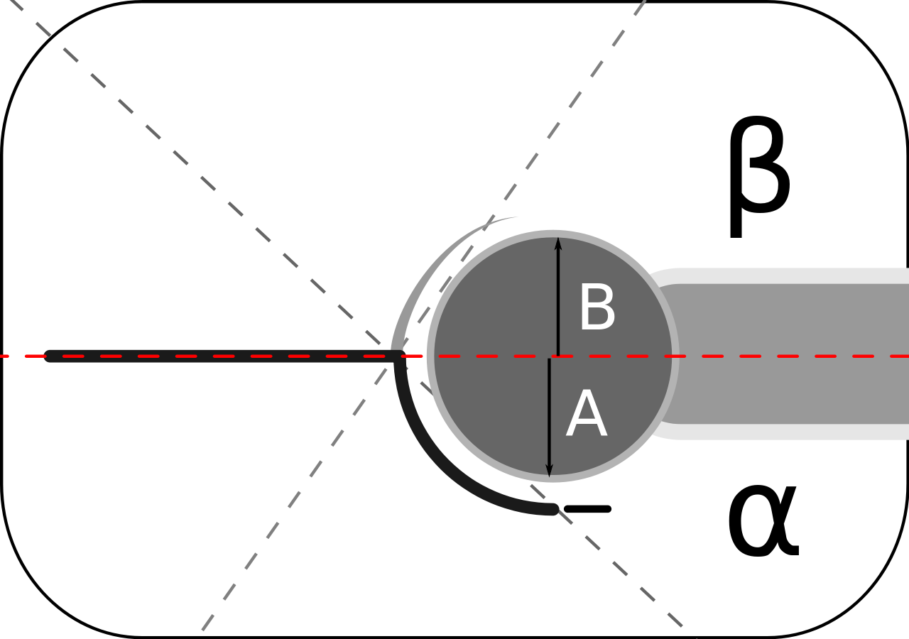
English: Lines and angles used in evaluation of hip dysplasia in Graf method which is the widely used throughout Europe. A Coronal ultrasound section obtained through the acetabulum.
Red:Baseline α: angle of bony acetabular roof with baseline, β angle of cartilaginous acetabular roof with baseline. Decreased α and increased β are associated with higher risk of hip dysplasia. Morin adapted the Graf technique to evaluate the percentage of the femoral head covered by the bony acetabulum. A/B*100: A: Portion of femur head covered by bone B: portion of femur head covered by cartilage. |
||
| Date | |||
| Source | Own work | ||
| Author | Nevit Dilmen (talk) | ||
| Permission (Reusing this file) |
I, the copyright holder of this work, hereby publish it under the following licenses: This file is licensed under the Creative Commons Attribution-Share Alike 3.0 Unported license. Attribution: © Nevit Dilmen
You may select the license of your choice. |
||
| Other versions |
 |


| Annotations | This image is annotated: View the annotations at Commons |
File history
Click on a date/time to view the file as it appeared at that time.
| Date/Time | Thumbnail | Dimensions | User | Comment | |
|---|---|---|---|---|---|
| current | 10:12, 29 March 2012 | | 725 × 510 (9 KB) | Nevit (talk | contribs) | update |
| 05:33, 28 March 2012 |  | 725 × 510 (8 KB) | Nevit (talk | contribs) | {{Information |Description={{en|Lines and angles used in evaluation of hip dysplasia}} |Source={{own}} |Date=2012 |Author= ~~~ |Permission={{User:Nevit/Not-PD}} |other_versions= }} Category:Hip dysplasia Category:Nevit Dilmen SVG |
You cannot overwrite this file.
File usage on Commons
The following page uses this file:
File usage on other wikis
The following other wikis use this file:
- Usage on de.wikipedia.org
- Usage on en.wikipedia.org
- Usage on hy.wikipedia.org
- Usage on sr.wikipedia.org
- Usage on tr.wikipedia.org
- Usage on uk.wikipedia.org